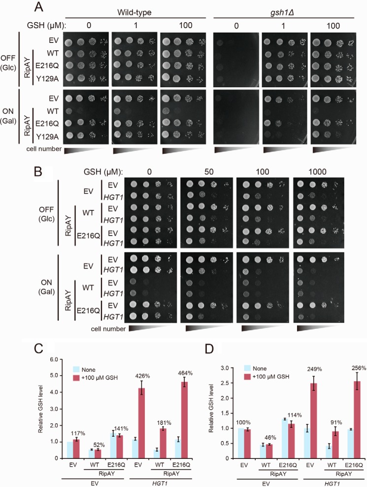
FIGURE 3.
Increased uptake of GSH does not rescue the growth inhibition effect caused by expression of RipAY. A, yeast wild-type and gsh1Δ/gsh1Δ homozygote cells carrying the indicated plasmids were spotted on SD (−Ura) (OFF) or SGal (−Leu, −Ura) (ON) plates supplemented with the indicated concentration of GSH. EV, empty vector. Cells were incubated at 26 °C for 2 days for SD (−Leu, −Ura) or 3 days for SGal (−Leu, −Ura). B, yeast cells carrying the indicated plasmids were spotted on SD (−Leu, −Ura) (RipAY expression: OFF) or SGal (−Leu, −Ura) (RipAY expression: ON) plates supplemented with the indicated concentration of GSH. Cells were incubated at 26 °C for 2 days for SD (−Leu, −Ura) or 3 days for SGal (−Leu, −Ura). RipAY or HGT1 were expressed under the control of a galactose-inducible GAL1 promoter or a strong constitutive TEF1 promoter, respectively. C, yeast cells carrying the indicated plasmids growing exponentially in SD (−Leu, −Ura) liquid medium were transferred to SGal (−Leu, −Ura) liquid medium and cultured at 26 °C for 19 h, and then GSH (100 μm) was supplemented at 30 min before termination of the culture. D, yeast cells carrying indicated plasmids growing exponentially in SD (−Leu, −Ura) liquid medium were transferred to SGal (−Leu, −Ura) liquid medium supplemented with or without 100 μm GSH and cultured at 26 °C for 19 h. The total GSH levels of the cell lysates from yeast cells expressing indicated proteins were measured. Values represent the mean ± S.E. (error bars) (n = 4 for C and n = 3 for D).

