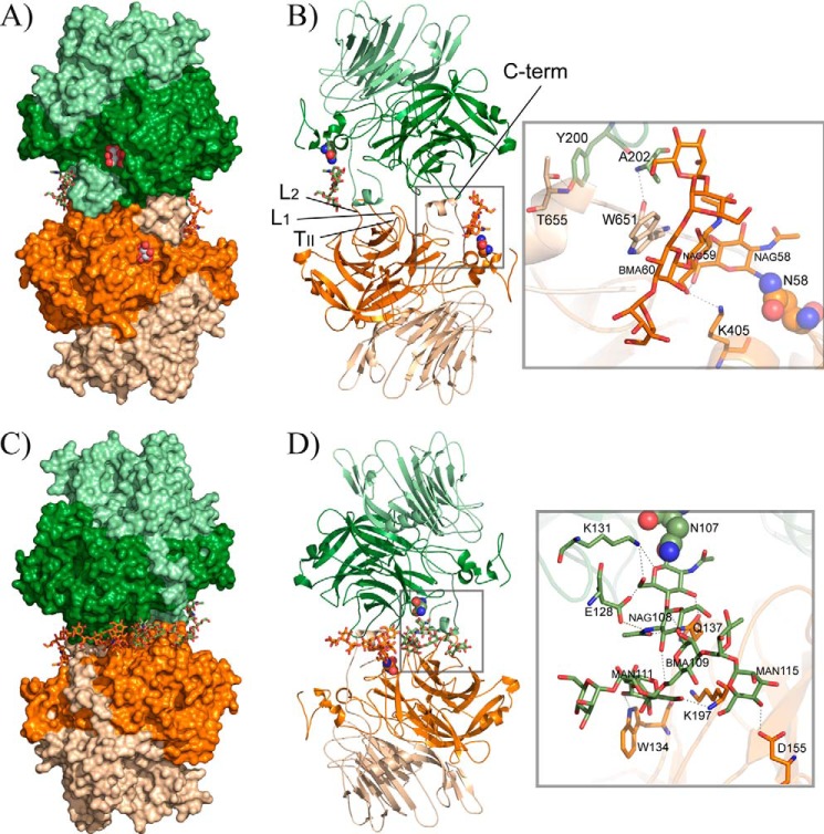
FIGURE 3.
XdINV dimer. A, molecular surface view of the dimer that is made by association of two subunits through their catalytic domains (dark orange/green) and the C-terminal region of the corresponding β-sandwich domains (light orange/green). A glycerol molecule found in the native protein crystals is shown in sphere representation at each active site pocket. The glycan chain is shown as sticks. B, same view of the dimer in schematic representation, highlighting the regions involved in the interface. Inset, a zoom showing the detailed intermolecular atomic interaction found around the glycan chain attached to Asn-58. C and D the opposite view of A and B showing the dimer and the atomic interactions at the interface around the glycan chain attached to Asn-107. NAG, GlcNAc; MAN, α-mannose; BMA, β-mannose.

