FIGURE 1.
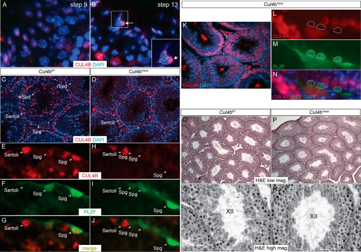
Germ cell-specific deletion of Cul4b does not affect testis morphology. A–D, IF of CUL4B in adult mouse testis. A and B, in wild type testis during spermiogenesis, CUL4B protein is dispersedly distributed in round spermatids (A), whereas more concentrated localization to the cytoplasmic lobe is detected in elongated spermatids (B). The developmental steps of spermatid differentiation were determined according to Russell et al. (47). C, CUL4B protein was detected in Sertoli cells (cells lining the seminiferous tubules with irregularly shaped nuclei and tripartite nucleoli), spermatogonia (Spg, cells lining the seminiferous tubules with round nuclei and often one visible nucleolus), round spermatids (rSpd, haploid cells located closer to the tubular lumen with smaller round nuclei and a large densely stained nucleolus), and elongated spermatids (eSpd, haploid cells located toward the tubular lumen with elongated nuclei) in a 4-month-old control Cul4bf/Y testis. D, Cul4b deletion through Vasa-Cre (Cul4bVasa) showed complete absence of the protein in all germ cell lineages, without affecting its expression in Sertoli cells. E–J, double IF of CUL4B and spermatogonial marker PLZF. CUL4B protein was readily detected in the PLZF-positive spermatogonia in the control testis (E–G) but not in the mutant testis (H–J). K–N, double IF of CUL4A and PLZF in Cul4bVasa testis showing no ectopic CUL4A expression in the absence of Cul4b. O–R, H&E staining showed normal histology of the Cul4bVasa testis, containing seminiferous tubules at all stages. K and L showed stage XII tubules of the two genotypes at higher magnification.

