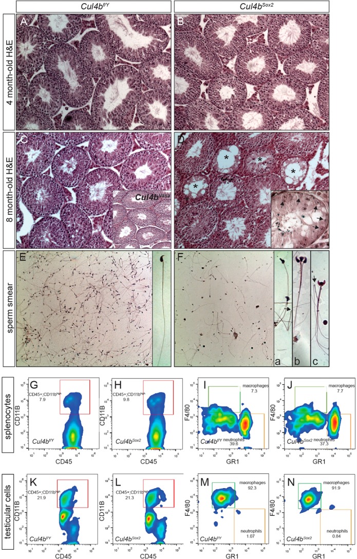
FIGURE 5.
Germ cell depletion in aged Cul4bSox2 testes. A–D, H&E staining of testis sections showing normal morphology at 4 months of age (B), but deteriorated structure by 8 months, with many empty seminiferous tubules (asterisks in D). The phenotype varied among individuals, with some having many tubules composed of Sertoli cells only (arrows, inset in D). Inset in C showed the normal histological organization of the 8-month-old Cul4bVasa testis. E and F, H&E staining of sperm smears of 8-month-old mice, showing reduced number of sperms in the mutant (F) with defective morphology. Inset a, deformed sperm head; inset b, triple head; inset c, double head. Arrows in insets a and c point to severed sperms with only flagella remaining. G–N, cell surface marker staining followed by FACS revealed no distinctive difference in macrophage population between the two genotypes.

