
FIGURE 1.
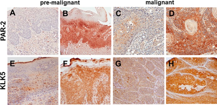
Immunohistochemical analysis of PAR-2 and KLK5 expression in pre-malignant oral lesions and OSCC. A and B, PAR-2 expression in pre-malignant oral lesions. A, 24% of lesions exhibited negative or low PAR-2 expression; B, 76% demonstrated moderate to high PAR-2 expression. PAR-2 antibody was used at a 1:50 dilution. Staining was detected using an avidin-biotin horseradish peroxidase system. C and D, PAR-2 expression in oral cancer. C, 24% of lesions exhibited negative or low PAR-2 expression; D, 76% demonstrated moderate to high PAR-2 expression. Antibody dilutions as in A. E and F, KLK5 expression in pre-malignant lesions. E, 37% of lesions exhibited negative or low KLK5 expression; F, 63% demonstrated moderate to high KLK5 expression. KLK5 antibody was used at a 1:50 dilution. G and H, KLK5 expression in oral cancer. Note that KLK5 expression in OSCC was previously published in Pettus et al. (17). In that study, 22% of lesions exhibited negative or low KLK5 expression, with 78% staining positive or strong positive. Magnification, ×200.