
FIGURE 1.
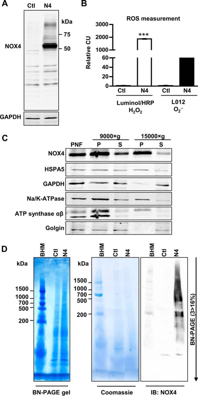
Characterization, differential centrifugation, and BN-PAGE of HEK293 cells lentiviral transduced with NOX4. A, representative Western blot for NOX4 in HEK293 cells (Ctl) and lentiviral-transduced NOX4-HEK293 cells (N4). B, luminol/HRP and L012 chemiluminescence assay in intact Ctl or N4-HEK293 cells normalized to Ctl cells (CU, chemiluminescence unit). n ≥ 3, mean ± S.D., *, p < 0.05; **, p < 0.01; ***, p < 0.001 relative to the corresponding Ctl HEK293 values. C, NOX4-HEK293 were mechanically homogenized and subcellular components were separated by differential centrifugation at 3,000 × g (PNF: postnuclear fraction), 9,000 × g and 15,000 × g to gain a pellet (P) and supernatant (S). D, 15,000 × g membrane pellets were solubilized with digitonin (6 g/g of protein) and used for BN-PAGE (3 > 16% acrylic amide); bovine heart mitochondria (BHM) served as weight standard. BN-PAGE gels were native blotted on a PVDF membrane and subsequently stained with Coomassie Blue and immunoblotted with anti-NOX4 antibody.