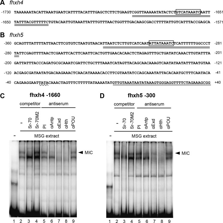
FIGURE 7.
A and B, sequence of the upstream region of fhxh4 (A) and fhxh5 (B). The boxed region indicates the hypothetical MIC binding site, the wavy line indicates the putative TATA box, the underlined sequence indicates the transcribed region, and the double underlined sequence indicates the oligonucleotide utilized for the EMSA analysis (fhxh4 −1660 and fhxh5 −300). C and D, binding of MIC to the upstream region of fhxh4 (C) and fhxh5 (D). The band that represents the presumptive MIC binding is indicated with the arrowhead. The binding was abolished by the non-labeled Sr-70 probe but not by the Sr-70M2 probe (lanes 3 and 4). Interference with the binding was also apparent after addition of antisera against Antp, Extradenticle (Exd), and Homothorax (Hth) but not after addition of anti-POU-M1 (POU) serum or preimmune (PI) serum (lanes 5–9).

