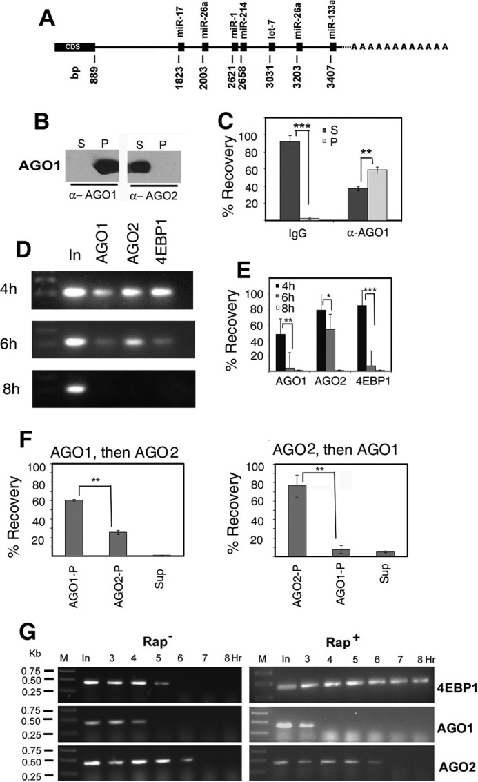
FIGURE 2.

Association of AGO isoforms with Ccnd1 mRNA. A, putative target sights for microRNAs on the 3′ UTR of Ccnd1 mRNA. The positions of the miRNA binding sites (with respect to the AUG start codon at +1) are indicated. cds, coding sequence. B and C, specificity of anti-AGO antibodies. B, an extract from 4-h stimulated L6 cells was treated with protein A-Sepharose beads coated with anti (α) -AGO1 or -AGO2 antibody and after centrifugation, equivalent amounts (4 μl) of the pellet (P) and supernatant (S) were analyzed by Western blot probed with anti-AGO1 or anti-AGO2 antibody. C, 4 h extract was incubated with Sepharose beads coated non-immune IgG (left) or anti-AGO1 antibody (right); RNA was prepared from the pellet (P) or supernatant (S) and equivalent amounts quantified by real time RT PCR as percent input recovered. In this and other figures, p values are indicated by stars: *, p < 0.5; **, p < 0.05; ***, p < 0.005. D and E, cells were serum stimulated for indicated times, and the lysates were subjected to R-CLIP using the specified antibodies. In, input RNA. D, agarose gel electrophoresis of the final PCR products. E, percent recovery calculated on the basis of Ct values by real time PCR (mean +S.D., n = 3–5). F, sequential immunoprecipitation of Ccnd1 mRNA from 4 h lysates and quantification of percent recovery in pellets (P) or final supernatant (S). Left, anti-AGO1 followed by anti-AGO2; right, the order of antibody was reversed. G, RT-PCR amplification of cross-linked and immunoprecipitated (CLIP) RNA from L6 myoblasts serum stimulated for the indicated times in the absence (left) or presence (right) of rapamycin, using antibodies against the proteins shown at the right. In, input RNA from pre-cleared lysate before immunoprecipitation. M, DNA markers of indicated size. Primers O-401 (forward) and O-402 (reverse; supplemental Table S2) were used to yield a 424-bp product.
