
FIGURE 8.
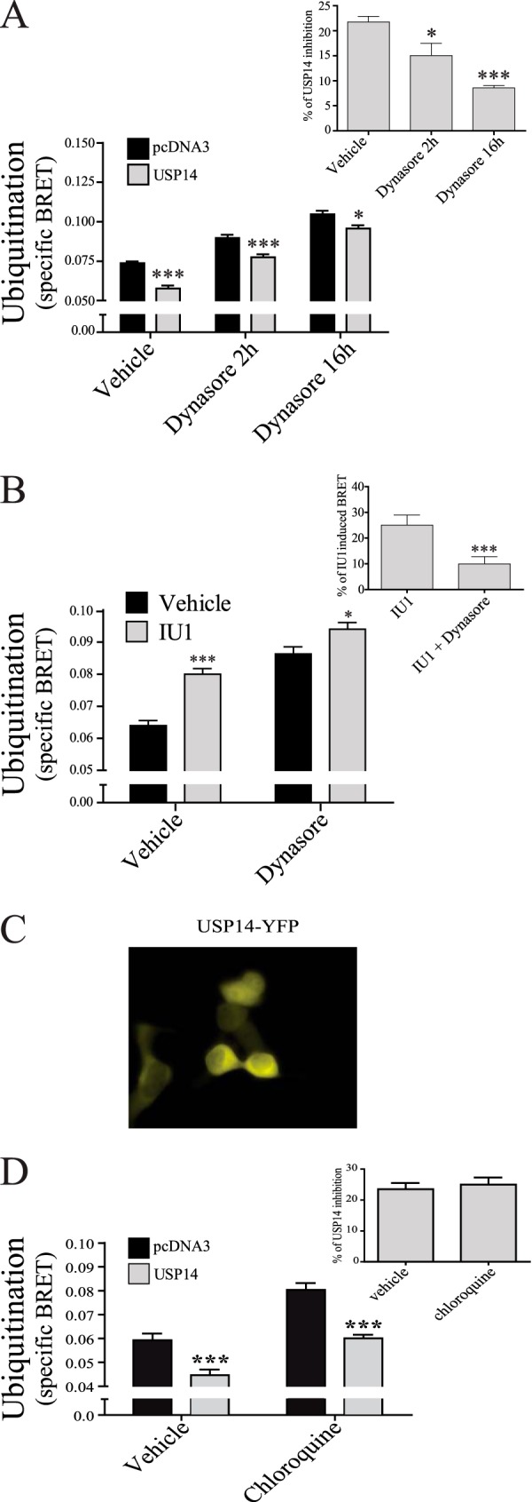
USP14 deubiquitination is decreased by internalization inhibitor Dynasore. HEK293T cells were transfected with Myc-GABAB(1b), HA-GABAB(2), MonoUbi-YFP or UbiAA-YFP, and either pcDNA3 or USP14. A, cells were treated or not (vehicle) for two or sixteen hours with 50 μm Dynasore and specific BRET ubiquitination signal calculated. Inset shows the percentage of inhibition of USP14 and calculated as in Fig. 5C. B, cells were treated or not (vehicle) for 2 h with 50 μm Dynasore, with or without IU1. Inset shows the percentage of IU1 induced BRET: IU1 treated minus vehicle dividing by vehicle condition. C, fluorescent microscopy illustrating the cytoplasmic distribution of USP14-YFP in COS7 cells co-expressing HA-GABAB(1a)-CFP, cMyc-GABAB(2). D, cells transfected as in A were treated or not (vehicle) for 2 h with 200 μm chloroquine and specific BRET ubiquitination signal calculated. Inset shows the percentage of inhibition of USP14 and calculated as in Fig. 5C. The results are presented as the mean ± S.E. of at least three independent experiments performed in triplicates. (*, p < 0.05; ***, p < 0.001.)