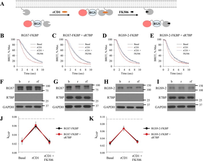
FIGURE 5.
Membrane orientation of RGS/Gβ5 does not impact GAP activity. A, schematic representation of reversible chemically-induced dimerization system with FKBP-binding partner on the C terminus of RGS (mCherry-RGS-FKBP). BRET assay was performed, and the deactivation phase during basal, rCD1, and rCD1 followed by FK506 treatment was plotted for: B, mCherry-RGS7-FKBP/Gβ5; C, mCherry-RGS7-FKBP/Gβ5 in the presence of sR7BP; D, mCherry-RGS9–2-FKBP/Gβ5; E, mCherry-RGS9–2-FKBP/Gβ5 in the presence of sR7BP. F–I, Western blot analysis was used to determine expression levels of RGS and R7BP. J–K, kGAP quantification was calculated as described above. Dotted line shows maximum kGAP value observed in this system when higher amount of RGS were used in the transfection. Each BRET trace and Western blot is from a single experiment representative of three independent experiments.

