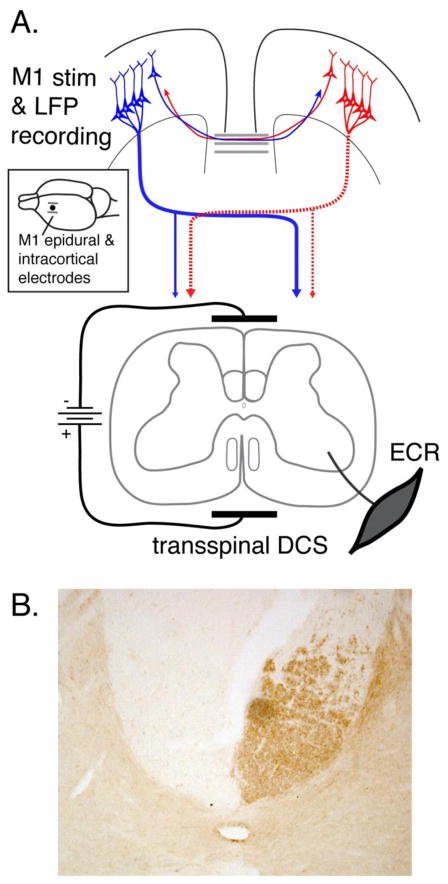
Figure 1.
A. Schematic of experimental setup. A bipolar epidural cortical stimulating electrode (see inset) was implanted over the fore limb representation of caudal M1. Recording electrodes (inset; single dot) were implanted between the two stimulating electrodes. tsDCS was delivered through patch electrodes with cathode placed dorsally over the C4 to T1 vertebrae and the anode placed over chest. EMG activity was recorded from the ECR muscle contralaterally. The corticospinal system on the left side of the figure (dotted) highlights that after PTX all CST projections from one hemisphere are eliminated. For the affected contralateral side, the only innervation is the spared ipsilateral CST. B. PKCγ staining marks CST axons in the dorsal column. The micrograph is from a representative PTX animal showing unilateral PKCγ loss due to the lesion.

