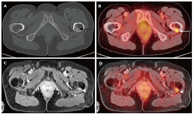
Figure 4.

A 59-year-old female with history of breast cancer status post resection and chemoradiation presents with left hip pain. Noncontrast CT of the pelvis (A) was unremarkable. PET/CT revealed increased focus of FDG uptake within the proximal left femur. Contrast-enhanced pelvic MRI (C) and PET-MRI (D) acquired simultaneously demonstrates abnormal nodular soft tissue mass within the proximal left femoral cortex with increased radiotracer uptake compatible with metastasis in this patient with breast carcinoma. PET: Positron emission tomography; MRI: Magnetic resonance imaging; CT: Computed tomography; FDG: 18F-fluorodeoxyglucose.
