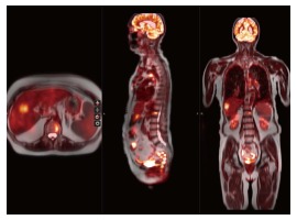
Figure 5.

A 66-year-old female with history of breast cancer status post resection and chemoradiation presents with chest pain. Whole-body PET-MRI reveal multiple abnormal nodular soft tissue masses within the lungs, mediastinum, ribs, liver and lumbar spine with increased FDG uptake consistent with metastasis in this patient with breast carcinoma. PET: Positron emission tomography; MRI: Magnetic resonance imaging; FDG: 18F-fluorodeoxyglucose.
