
Figure 4.
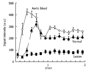
2D magnetic resonance perfusion demonstrates severely hypoperfused ablated lesions after magnetic resonance-guided high intensity focused ultrasound compared to remote non-ablated tissue. Diamond: Aortic blood; Triangle: Viable renal tissue; Square: Ablated lesion. a.u.: Arbitrary units.