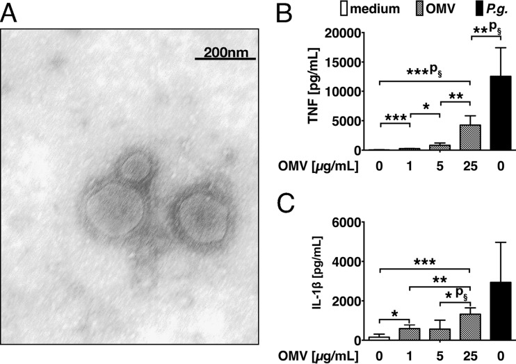
FIG 1.
Secretion of proinflammatory cytokines in response to P. gingivalis OMV. (A) Trans-electron microscopy of the purified OMV. The image shows a negative stain with 2% methylamine tungstate and is representative of the results of n = 6 experiments. (B and C) Human CD14+ monocytes were stimulated with OMV at the indicated concentrations or with live P. gingivalis (P.g.). Cellular supernatants were harvested after 24 h and cytokines quantified by ELISA. (B) TNF. (C) IL-1β. The graphs summarize the results obtained from n = 6 independent donors (n = 3 independent experiments). ***, P = 0.0002; ***p§ = 0.0008; *, P = 0.0281; **, P = 0.0031; **p§ = 0.0027 (B); *, P = 0.0119; ***, P = 0.0003; **, P = 0.0051; *p§ = 0.0217 (C).

