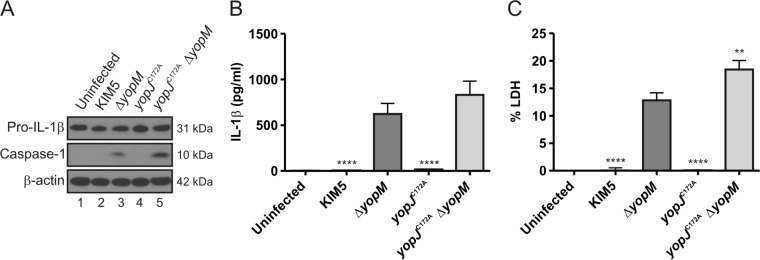
FIG 6.
Measurement of processed caspase-1, pro-IL-1β, mature IL-1β, and pyroptosis in BMDMs infected with Y. pestis. BMDMs from C57BL/6 mice were primed with 100 ng/ml LPS for 18 h and left uninfected or infected with KIM5, KIM5 ΔyopM, KIM5 yopJC172A, or KIM5 yopJC172AΔyopM at an MOI of 30. (A) At 90 min postinfection, lysates were collected, processed, and subjected to Western blotting using antibodies against pro-IL-1β and p10, the 10-kDa subunit of caspase-1. β-Actin was blotted as a loading control. Quantification of the processed caspase-1 bands showed that the signal for KIM5 yopJC172AΔyopM was 2.3 times greater than that for KIM5 ΔyopM. At 90 min postinfection, supernatants were collected, and secreted IL-1β was measured by ELISA (B); pyroptosis was quantified as percent LDH release (C). The data in panel B and C represent average values ± the standard error of the means from three independent experiments. **, P < 0.01; ****, P < 0.0001, as determined by one-way analysis of variance compared to results for KIM5 ΔyopM-infected macrophages.

