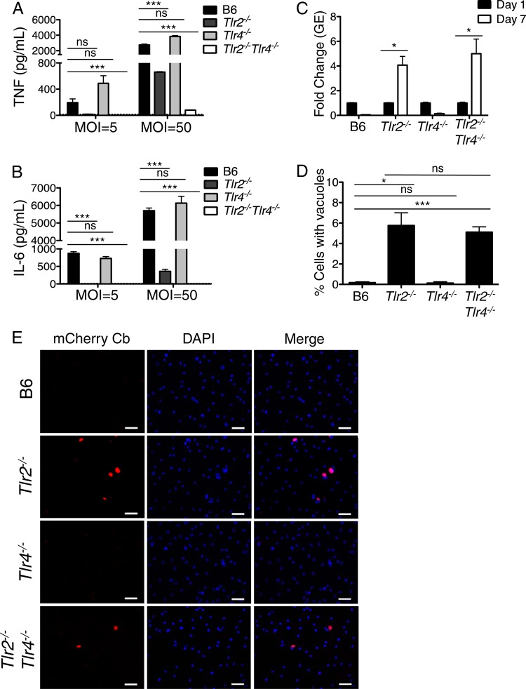
FIG 1.
TLR2 and TLR4 mediate immune responses to Coxiella burnetii Nine Mile II in C57BL/6 macrophages. (A and B) C57BL/6, Tlr2−/−, Tlr4−/−, and Tlr2−/− Tlr4−/− bone marrow-derived macrophages (BMDMs) were infected with WT C. burnetii NMII at an MOI of 5 or 50 for 24 h. Levels of TNF and IL-6 in the supernatants were measured by ELISA. Graphs show the means ± standard errors of the means (SEM) from triplicate wells. Results are representative of three independent experiments. (C) C57BL/6, Tlr2−/−, Tlr4−/−, and Tlr2−/− Tlr4−/− BMDMs were infected with WT C. burnetii NMII at an MOI of 100. At days 1 and 7 postinfection, bacterial uptake and replication were measured as genomic equivalents (GEs) by qPCR. Graphs show the fold change in GEs on day 7 relative to GEs on day 1 ± SEM from triplicate wells. (D) Seven days postinfection, BMDMs of the indicated genotypes infected with mCherry-expressing WT C. burnetii NMII at an MOI of 100 were fixed, stained with DAPI, and examined by fluorescence microscopy. The number of large mCherry-expressing NMII-containing vacuoles greater than 5 μm in size was determined and calculated as a percentage of the total cell number. Graphs show the mean percentage of cells containing large NMII vacuoles ± SEM from triplicate coverslips. At least 300 cells were counted per coverslip. Results are representative of two independent experiments. (E) Representative fluorescence micrographs of mouse BMDMs of the indicated genotypes infected with mCherry-expressing C. burnetii (Cb) NMII at an MOI of 100 and fixed and stained with DAPI on day 7 postinfection. Images were taken at ×40 magnification. Scale bars represent 25 μm. *, P < 0.05; **, P < 0.01; ***, P < 0.001; ns, no significance.

