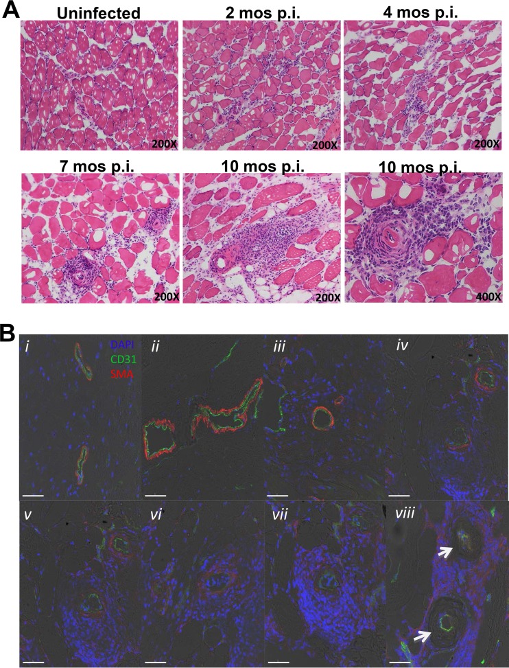
FIG 2.
Histopathology of systemic necrotizing vasculitis lesions in skeletal muscle late after T. cruzi infection of C57BL/6J mice. Sections of quadriceps muscles were analyzed. (A) Time course of infection. Samples were obtained from mice at the indicated time points after infection and stained with H&E. Images are representative of a total of 3 mice analyzed per time point. (B) Development of fibrinoid necrosis. Samples of vascular lesions with different degrees of severity (i to viii) were stained for immunofluorescence with DAPI (blue) and with antibodies directed against CD31 (green) and smooth muscle cell actin (SMA) (red). Images are representative of 3 mice analyzed 10 months after infection. Confocal immunofluorescence images are superimposed on bright-field images. Arrows, absence of smooth muscle actin.

