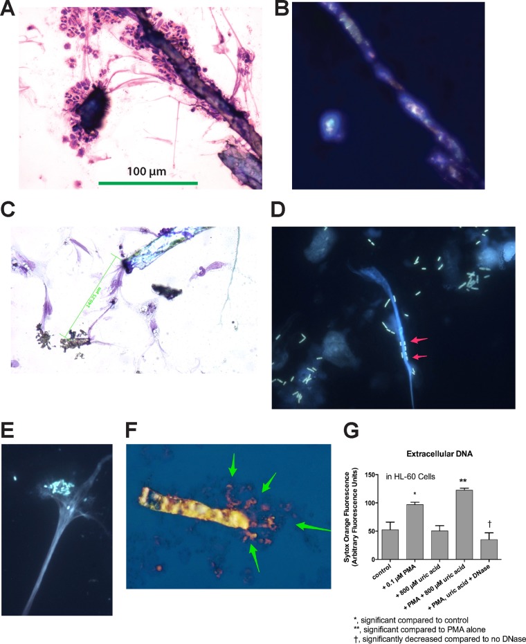
FIG 3.
Incorporation of uric acid into DNA NETs. (A, B, and F) Formation of DNA NETs from rabbit peripheral blood leukocytes stimulated with 0.1 μM PMA. (C to E) Images obtained with differentiated HL-60 cells. (A) Bright-field microscopy of PMA-stimulated PBLs showing adherence of leukocytes to crystals and stringy filaments connecting the crystals (three-step stain; magnification, ×100). (B) Polarization microscope image of the same field as in panel A, demonstrating that the crystals show birefringence. The 100-μm scale bar in panel A also applies to panel B. (C) Bright-field microscope image showing that crystals are connected over long distances by stringy filaments, which appear to originate from the nuclei of PMA-treated HL-60 cells. (D) Fluorescent DH5α bacteria expressing green fluorescent protein (GFP) adhere to DNA NETs (arrows) (magnification, ×600; Hoechst stain for DNA). (E) Adherence of human EPEC E2348/69 bacteria to DNA NETs generated from PMA-stimulated HL-60 cells stained with Hoechst dye for DNA. Note that the EPEC bacteria demonstrate their typical localized adherence pattern even when adhering to DNA (magnification, ×600). (F) Merged image showing a birefringent uric acid crystal (photographed by polarization microscopy) superimposed on the same field photographed under fluorescence, with staining using 10 μM Sytox Orange. The orange-stained DNA seems to emanate from one end of the uric acid crystal (arrows). The uric acid crystal itself is 118 μM in length. (G) Uric acid-induced DNA release from HL-60 cells at 3 h of treatment. The error bars indicate SD.

