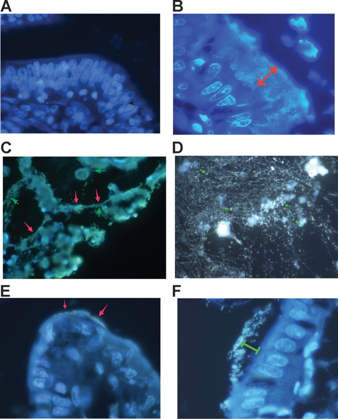
FIG 4.
Formation of DNA NETs in vivo in the lumen of the GI tract in rabbit. (A) Hoechst stain of the ileum of a healthy uninfected rabbit. The intestinal segment was fixed in formaldehyde, embedded in paraffin, sectioned, and then deparaffinized and stained with 5 μg/ml Hoechst 33258 dye. (B) Hoechst stain of the ileum of a rabbit intestinal segment infected with rabbit STEC strain E22-stx2 for 20 h. The lumen of the gut is at the top right. STEC bacteria are adhering as a thick biofilm (double-headed arrow, which is 20.8 μm in length). The scale in panel B also applies to panels A, C, and D. (C) DNA NETs formed in vivo from rabbit intestine infected with rabbit EPEC strain E22. Cells that are being sloughed into the intestine are connected by DNA strands (red arrows) and surrounded by bacteria. A few individual EPEC bacteria are indicated by green arrows (magnification, ×600). (D) Another field from an intestine infected with EPEC E22 and stained with 10 μg/ml DAPI, showing that the DNA NETs can form a delicate, lacy network and not just linear strands. A few individual EPEC bacteria are again indicated by arrows. (E and F) Effect of DNase I on NETs formed during infection with EPEC E22 (magnification, ×1,000). (E) DNase-treated intestinal loops showed markedly fewer adherent bacteria; the arrows indicate a few E22 bacteria adhering to a villus tip. (F) Remnant bacteria observed in DNase-treated intestinal loops seemed to be adhering “at a distance” rather than in the typical intimate adherence pattern seen in panel B. Green bracket, 8 μm.

