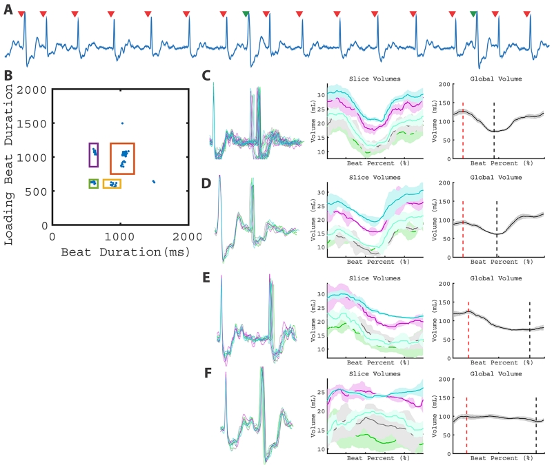
Figure 6.
LV function with interpolated PVCs. The ECG (A) and 2D plot (B) depict 4 beat types: sinus rhythm (C), post-PVC sinus (D), interrupted sinus (E), and the interpolated PVC contractions (F). Global volume quantification suggests interpolated PVCs affect post-PVC loading and do not result in substantial stroke volume. Global volume measurements are found in Table 1 (Patient 8).

