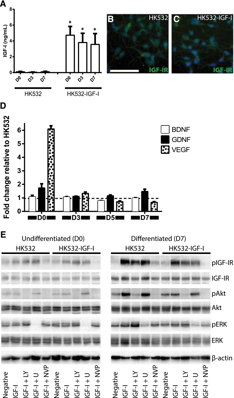
Figure 1.
IGF-I production, growth factor profile, and signaling in HK532 and HK532-IGF-I cells. (A): Production of IGF-I in HK532 and HK532-IGF-I throughout early differentiation. Representative immunocytochemistry images of D7 HK532 (B) and HK532-IGF-I (C) cells labeled with 4′,6-diamidino-2-phenylindole (DAPI) (blue) and IGF-IR (green). (D): BDNF, GDNF, and VEGF production in undifferentiated HK532 and HK532-IGF-I cells (D0) and throughout early differentiation (D3, D5, and D7). Growth factor production is expressed as fold change relative to parental HK532 cells. (E): Western blot analysis of IGF-I signaling in undifferentiated and differentiated (D7) HK532 and HK532-IGF-I cells. Cells were treated with an inhibitor panel of LY, U, or NVP for 1 hour, followed by IGF-I treatment for 30 minutes. All blots were probed with pIGF-IR, IGF-IR, pERK, ERK, pAKT, and AKT. β-actin was used as a loading control. Data are presented as mean + SD or are representative images of at least three independent experiments. Scale bar = 50 μm. ∗, p < .05. Abbreviations: BDNF, brain-derived neurotrophic factor; D0 (D3, D7), day 0 (day 3, day 7); GDNF, glial cell line-derived neurotrophic factor; IGF-I, insulin-like growth factor-I; IGF-IR, insulin-like growth factor-I receptor; LY, LY294002; NVP, NVPAEW541; U, U0126; VEGF, vascular endothelial growth factor.

