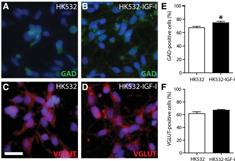
Figure 4.
Terminal phenotype of HK532 and HK532-IGF-I cells. (A, B): Representative immunocytochemistry (ICC) images of D7 HK532 and HK532-IGF-I cells labeled with 4′,6-diamidino-2-phenylindole (DAPI) (blue) and GAD65 (green). (C, D): Representative ICC image of D7 cells labeled with DAPI (blue) and VGLUT (red). (E): Quantification of GAD65-positive gamma-aminobutyric acid (GABA)ergic neurons in HK532 and HK532-IGF-I cells. HK532-IGF-I cells preferentially differentiate into GABAergic neurons (∗, p < .05 vs. HK532). (F): Quantification of VGLUT-positive glutamatergic neurons in HK532 and HK532-IGF-I cells. Data are presented as representative images or mean + SD of at least three independent experiments. Scale bar = 200 µm. Abbreviations: D7, day 7; GAD, glutamic acid decarboxylase; VGLUT, vesicular glutamate transporter.

