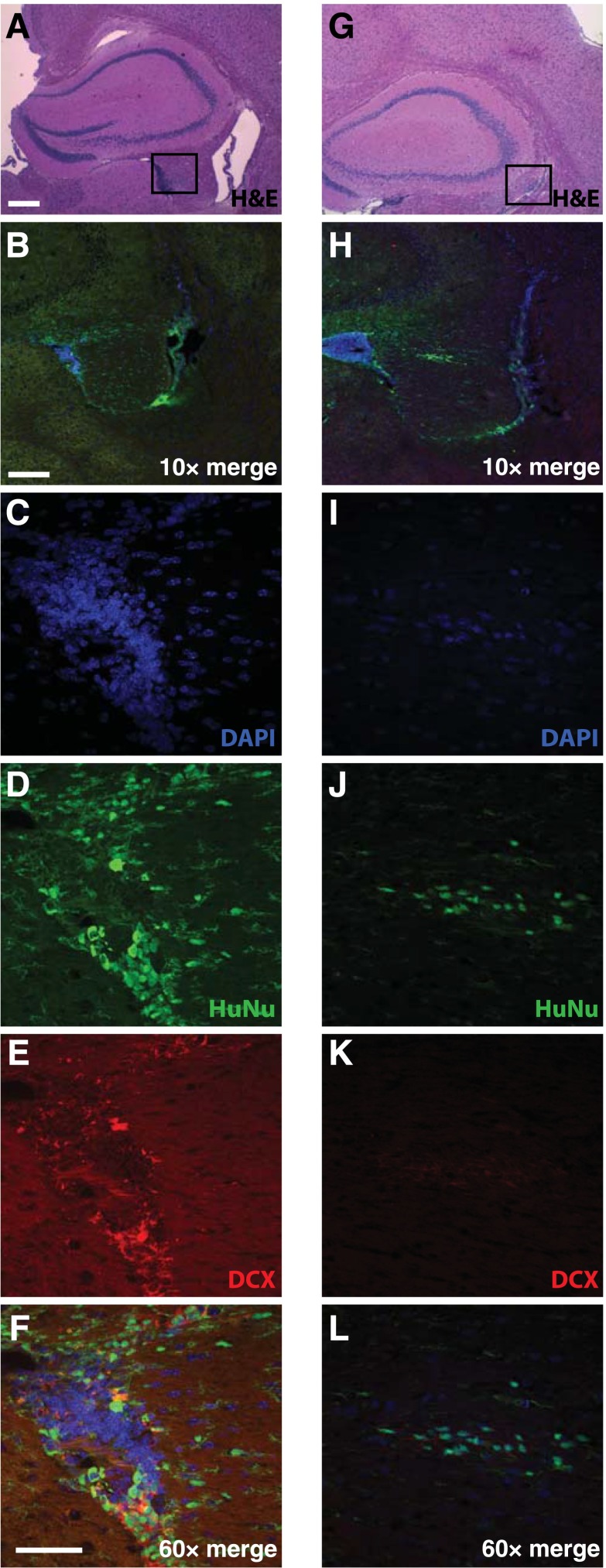
Figure 6.
HK532-IGF-I cells survive grafting into APP/PS1 mice with Alzheimer’s disease (AD) and wild-type (WT) mice. Representative images of HK532-IGF-I cells in the hippocampal area of APP/PS1 AD (A–F) and WT (G–L) mice 10 weeks following transplantation to the fimbria fornix. H&E staining shows the transplanted target area (square) in AD (A) and WT (G) mice. Immunofluorescent DAPI, HuNu, and DCX labeling of human early neural precursor cells in the hippocampal area of AD (B–F) and WT animals (H–L). Data are presented as representative images (A–F: APP/PS1, n = 4; G–L: WT, n = 4). (A, G): Scale bar = 200 µm. (B, H): ×10 scale bar = 200 µm. (C–F, I–L): ×60 scale bar = 50 µm. Abbreviations: DAPI, 4′,6-diamidino-2-phenylindole; DCX, Doublecortin; H&E, hematoxylin and eosin; HuNu, human nuclei.

