Figure 2.
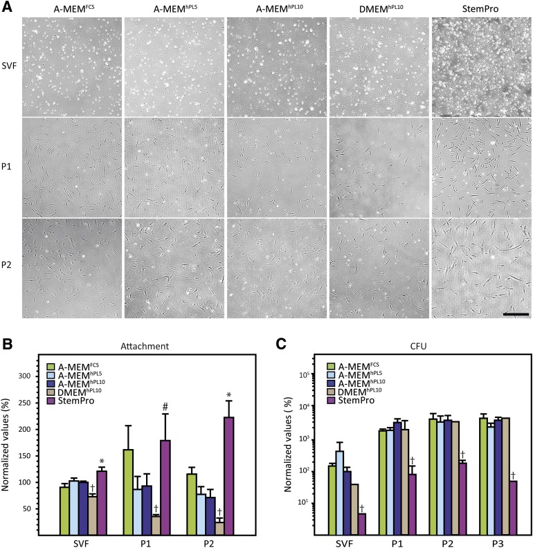
Cell morphology, attachment (A, B), and clonogenicity (C). (A): Differences in the degree of cell attachment were observed after seeding and overnight incubation. Additionally, morphologic differences were seen between passages. (B): Quantification of cell attachment revealed significant differences between media. (C): Colony-forming potential of the different media. It can be seen that StemPro significantly underperformed compared with the other media. The results are presented as mean ± SEM. Cell number was normalized to the cell number of each individual donor for A-MEMhPL10 at passage 1. †, Statistically different from all other media at p < .05. ∗, StemPro statistically different from all other media. p < .05. #, StemPro statistically different from all other media except A-MEMFCS at p < .05. Scale bar = 500 µm. Abbreviations: A-MEM, α-minimum essential medium; CFU, colony-forming unit; DMEM, Dulbecco’s modified Eagle’s medium; FCS, fetal calf serum; hPL5, 5% human platelet lysate; hPL10, 10% human platelet lysate; P, passage; SVF, stromal vascular fraction.

