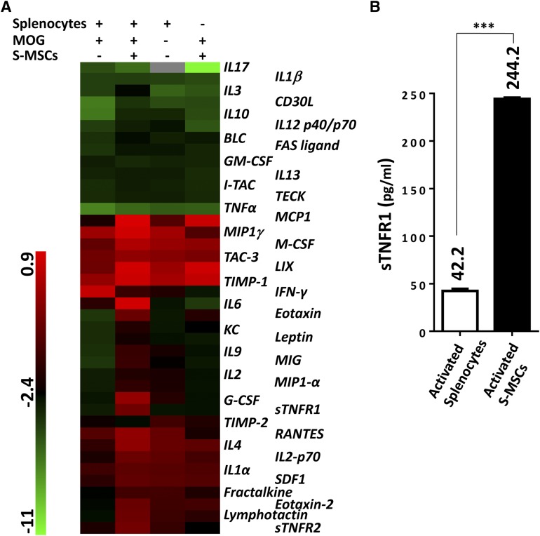
Figure 4.
High levels of sTNFR1 were secreted by S-MSCs. (A): Mouse cytokine antibody array screening of supernatant derived from cultured cells indicated on the top of the figure (six mice were used for each group). (B): Splenocytes restimulated with MOG35–55 in the presence of S-MSCs for 24 hours and then splenocytes and S-MSCs were separated and subsequently cultured for another 24 hours. The supernatants were harvested, and the expression of sTNFR1 was examined by enzyme-linked immunosorbent assay (n = 4). The amounts of secreted sTNFR1 were listed. ∗∗∗, p < .001. The data in (B) are representative of at least two independent experiments. Abbreviations: MOG, myelin oligodendrocyte glycoprotein; S-MSC, skin-derived mesenchymal stem cell; sTNFR1, soluble tumor necrosis factor receptor 1.

