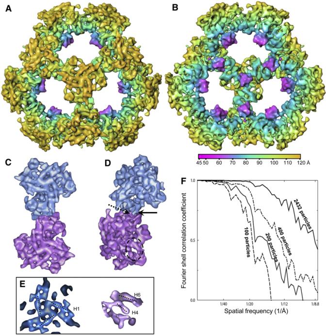
Figure 3. 3D Reconstruction of Human tE2 at 8.8 Å Resolution.
(A and B) Shaded, radially colored surface representation of (A) outside and (B) inside views of the human tE2 core along a three-fold axis. Secondary structure elements (cylindrical helices and globular β sheets) are discernable.
(C and D) (C) Top and (D) side views of two adjacent tE2 trimers extracted from the 3D reconstruction of the tE2 core, viewed along a two-fold axis. The solid and dashed arrows in (D) indicate the “ball” and “socket,” respectively, of the characteristic interaction between two adjacent trimers. The dashed oval marks a region with two helices, which is blown up in the right panel of (E).
(E) Close-ups of the 3D map of human tE2, showing some rod-shaped helix densities. Left: top view of three H1 helices surrounding a three-fold axis. Right: side view of the circled area in (D) showing H4 and H6 helices (see helix numbering and details in Figures 4 and 6).
(F) Fourier shell correlation coefficient showing improvement in resolution as the number of particles increases.

