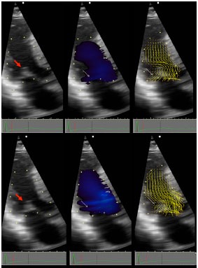
Fig. (1).

OHCM patient with ejection SAM. Vector Flow Mapping (VFM) is a novel method of processing Doppler information that demonstrates the vector of local blood flow velocity in intravascular structures. It differs from routine color Doppler: in VFM, a post-processing computational algorithm extracts information from the distribution of Doppler color flow in the beam direction and estimates the radial (perpendicular) component of the flow distribution, and displays it without angle dependence demonstrating direction and magnitude of blood flow velocity over 360 degrees. Local flow velocity is depicted as yellow lines proportional to, and in the direction of local velocity, indicated by red head of vector. Top panels are all pre-SAM frames; bottom panels are all post-SAM frames. Red arrow points to coapted MV. Early SAM is seen by comparing the 2D frames top and bottom left. White arrows point, in middle panels to blue color flow posterior to the MV, and in right panels to ricochet flow off the leaflet and into the cul-de-sac. Note that vector flow impacts the posterior surface of the mitral leaflets with high angle of attack and then ricochets off the leaflet into the cul-de-sac behind the valve. Neither blue flow posterior to leaflet, nor ricochet are seen in normals. Reproduced by permission from Ro R et al. Vector Flow Mapping in Obstructive Hypertrophic Cardiomyopathy to Assess the Relationship of Early Systolic Left Ventricular Flow and the Mitral Valve. J Am Coll Cardiol 2014; 64: 1984-95.
