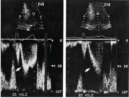
Fig. (4).

Comparison of left ventricular pulsed Doppler tracings before treatment (left) and after successful medical treatment (right) for patient 2. The sample volume was 2.5 cm apical of mitral valve coaptation point. Before treatment, ejection acceleration was rapid (arrowhead), and velocity peaked in the first half of systole. After treatment, ejection acceleration was slowed (arrowhead), and velocity peaked in the second half of systole. Systolic anterior mitral motion was delayed, and a 96 mm Hg gradient was eliminated. Note that although acceleration slowed, peak velocity remained virtually unchanged. This contrast highlights the importance of acceleration and the timing of ejection in successful medical therapy. The velocity calibration is identical in both panels. The scale is 20 cm/s between white marks. Reproduced by permission from Sherrid, M.V. et al. Mechanism of benefit of negative inotropes in obstructive hypertrophic cardiomyopathy. Circulation 1998; 97: 41-7.
