
Figure 1. YAP Overexpression Promotes the Generation of Human Naive ESCs.
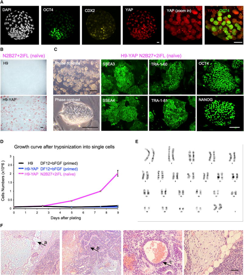
(A) YAP protein is localized to the nucleus in both the trophectoderm and the ICM of human blastocysts, as shown by immunofluorescence. White, DAPI; green, OCT4 (ICM); yellow, CDX2 (trophectoderm); red, YAP. Representative image of n = 16 human blastocysts. Scale bar, 20 μm.
(B) In naive medium N2B27+2iFL, human H9 ESCs with YAP overexpression (H9-YAP) maintain expression of the pluripotency marker AP, while control H9 cells differentiate and do not express AP. Scale bar, 500 μm.
(C) H9-YAP cells in N2B27+2iFL have a naive-specific dome-like colony morphology and show strong positive immunostaining for pluripotency markers SSEA3, SSEA4, TRA-1-60, TRA-1-81, OCT4, and NANOG. Black scale bar, 500 mm; white scale bar, 150 μm.
(D) Growth curves of H9 in DF12+bFGF primed medium, H9-YAP in DF12+bFGF primed medium, and H9-YAP in N2B27+2iFL naive medium after trypsinization to single cells. Only H9-YAP cells in naive medium have a high proliferate rate. Error bars represent SD.
(E) H9-YAP cells in N2B27+2iFL have a normal female karyotype (46, XX), as evaluated after 20 passages in naive culture conditions.
(F) H9-YAP cells in N2B27+2iFL are able to form teratomas comprising tissues derived from all three germ layers. (a) Adipocytes (mesoderm). (b) Neural tissue (ectoderm). (c) Epithelium (endoderm). White scale bar, 200 μm; black scale bar, 50 μm.