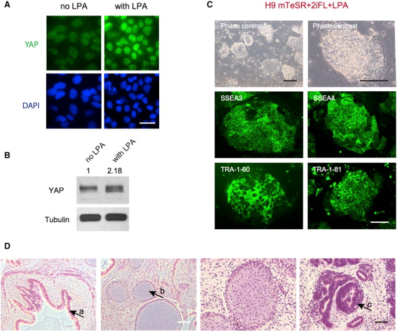
Figure 2. LPA Can Activate YAP and Promotes a Naive State of Pluripotency.

(A) LPA increases the level of YAP in human H9 ESCs, as shown by immunofluorescence. Immunostaining was performed 2 days after adding LPA. Green, YAP; blue, DAPI. Scale bar, 50 μm.
(B) Western blotting confirming that LPA increases YAP protein levels. Tubulin was used as loading control.
(C) H9 cells in N2B27+2iFL+LPA naive medium have a naive-specific dome-like colony morphology and show strong positive immunostaining for pluripotency markers SSEA3, SSEA4, TRA-1-60, and TRA-1-81. Black scale bar, 500 μm; white scale bar, 150 μm.
(D) H9-YAP cells in N2B27+2iFL are able to form teratomas comprising tissues derived from all three germ layers. (a) Gut-like epithelium (endoderm). (b) Cartilage (mesoderm). (c) Neural tissue (ectoderm). White scale bar, 200 μm; black scale bar, 50 μm.
