
Figure 6.
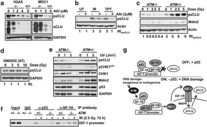
sCLU induction after DNA damage is mediated by ATM. (a) H2AX−/− and MDC1−/− cells were exposed to ATR kinase inhibitor (1–4 μM, 48 h) and harvested for immunoblotting. (b) MCF-7 cells were pretreated with ATR kinase inhibitor (2 μM, 1 h) before IR or TPT treatments, and then harvested at 72 h for western blotting. (c) Immortalized AT (ATM−/−) and AT reconstituted for ATM (ATM+/−) cells were exposed to 0–5 Gy and analyzed 48 h later by immunoblotting. (d) Primary human AT-deficient fibroblasts, GM2052, were treated with 0–10 Gy and analyzed 72 h later by immunoblotting. (e) ATM−/− and ATM+/− cells were exposed to UV (1 or 3 J/m2) or mock-treated, and harvested at 48 h for immunoblotting. (f) ATM−/− and ATM+/− were exposed to 2.5 Gy and harvested 72 h later for ChIP. p53 and NF-YA were immunoprecipitated, and the IGF-1 promoter was amplified using specific primers. (g) Model of ATM-IGF-1–MAPK–sCLU signaling pathway in the absence or presence of p53 and DNA damage. RL, relative levels.