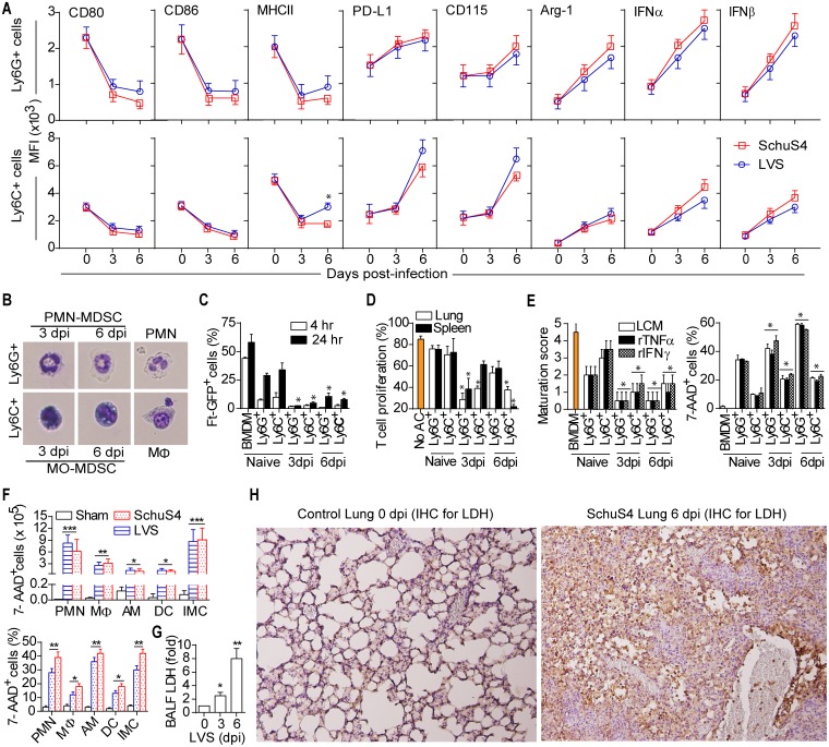
Fig 5. Ft-elicited myeloid cells are IMC/MDSC.
(A) MFI of the indicated myeloid cell marker expressed in Ly6G+ or Ly6C+ cells in lungs during Ft infection (mean ± SD of two (SchuS4, n = 6 mice) or three (LVS, n = 9 mice) independent experiments; MFI values for all the markers in Ft-infected mice at 3 and 6 dpi were significantly (p<0.05) different from control mice at 0 dpi. *p<0.05 indicates difference between SchuS4 and LVS). (B) Giemsa staining of isolated Ly6G+ or Ly6C+ cells from LVS-infected lungs. Note immature nuclear morphologies of ring/band-shaped (Ly6G+) or small round (Ly6C+) nuclei. (C) Percent Ly6G+ or Ly6C+ cells positive for phagocytosed Ft-GFP upon in vitro infection. (D) In vitro proliferation of T cells (CFSE dilution) with and without accessory cells (AC) such as Ly6G+ and Ly6C+ cell populations. (E) In vitro differentiation/maturation score and 7-AAD positivity for Ly6G+ or Ly6C+ cells and frequency of 7-AAD+ Ly6G+ and Ly6C+ cells cultured with indicated factors (For C-E, mean ± SD of two independent experiments, Student’s t-test, *p<0.05). (F) Numbers/frequency of 7-AAD+ myeloid cell subsets from Ft-infected lungs at 6 dpi (mean ± SD of two (SchuS4) or three (LVS, Sham) independent experiments, Student’s t-test, *p<0.05, **p<0.01, ***p<0.001). (G) LDH activity in BAL fluid following LVS-infection (mean ± SD of two independent experiments, Student’s t-test, *p<0.05, **p<0.01). (H) Positive immunoreaction for localization of LDH, as an indicator of necrosis, in lung sections from control or SchuS4-infected mice (IHC with hematoxylin counterstaining, 400x).

