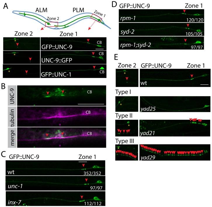
Fig 1. The cluster of UNC-9 puncta in PLM neurons represents gap junctions in vivo.
(A) Expression patterns of GFP::UNC-9 fusion proteins represent the localization of gap junctions in PLM neurons. At the top panel, a cartoon picture shows the morphology of PLM neurons. The red rectangles highlight two gap junction zones in PLM neurons. In bottom panels, images of expression patterns of GFP::UNC-9, UNC-9::GFP and GFP::UNC-1 in PLM neurons. Red arrowheads point to GFP puncta at gap junction zones. Yellow stars label ALM cell bodies. CB: PLM cell bodies. (B) Images of immunostaining results show that endogenous UNC-9 forms similar punctate structures at gap junction zone 1. UNC-9 was stained using Rabbit anti-UNC-9 antibody (green color). PLM neurons were labeled using mouse anti-acetylated tubulin antibody (pink color). We confirmed those neurons were PLM neurons based on their morphology and the position of cell bodies. CB: PLM cell bodies. (C) Representative images show that loss-of-function mutations in unc-1 or inx-7 does not affect GFP::UNC-9 puncta. (D) Suppression of chemical synapse formation by mutating rpm-1 and syd-2 does not affect GFP::UNC-9 puncta in PLM neurons. (E) Three types of mutants identified in the genetic screen for gap junctions. Red arrowheads point to GFP::UNC-9 puncta. Yellow stars label ALM cell bodies. In figures C and D, the number displayed at each image shows the number of animals with wild type GFP::UNC-9 puncta/ total animals. Scale bar: 10 μm. Detailed strain information of all figures is listed in the S1 Table.

