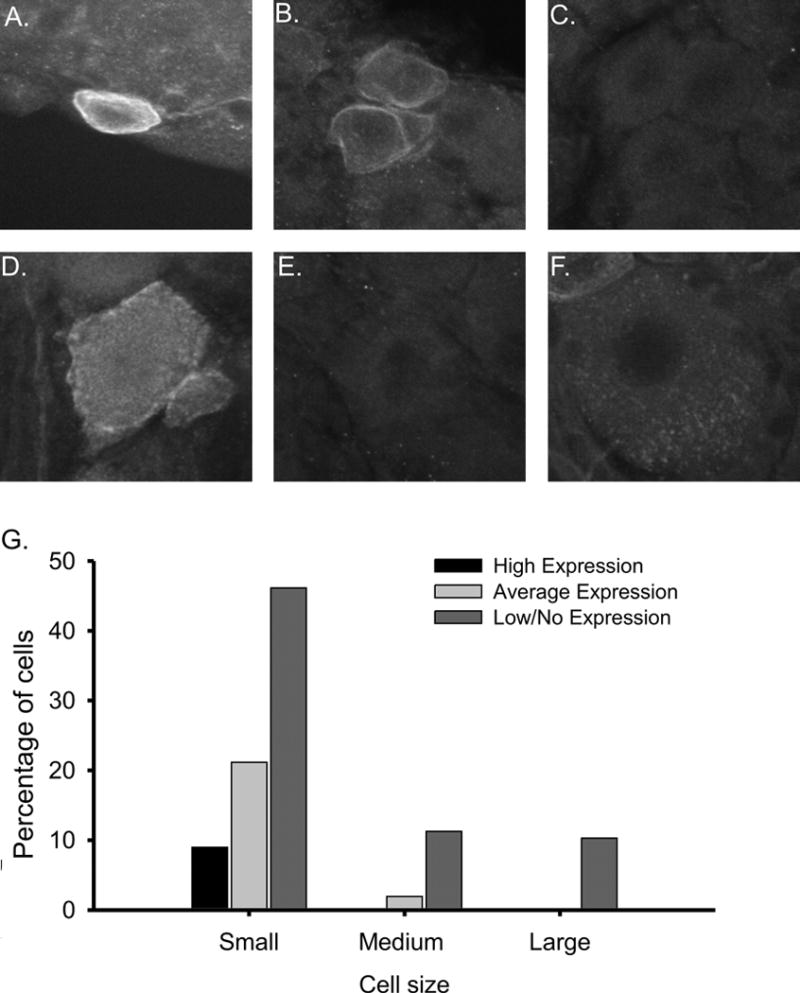
Figure 2. Characterization of levels of AKAP150 expression in rat DRG neurons using confocal microscipic analysis.

Scale bars = 50 μM. A. AKAP150 is highly expressed at the membrane of the soma and axon initial segment (AIS) in a subpopulation of small diameter DRG neurons morphologically resembling small-sized nociceptors. AKAP150 is expressed at the membrane of the soma and AIS of these neurons. B. Small diameter DRG neuron expressing moderate levels of AKAP150. C. A small DRG neuron displaying little to no AKAP150 expression. D. A small subset of medium diameter neurons expressed AKAP150, but the majority of medium and large diameter DRG neurons lacked any AKAP150 immunoreactivity (E, F). G. Histogram of cell area and AKAP150 intensity in small, medium and large DRG neurons. Cell size and AKAP150 intentiAKAP150 expression levels where highest in small diameter DRG neurons. Within the small diameter neurons, a subset displayed significantly higher levels of AKAP150.
