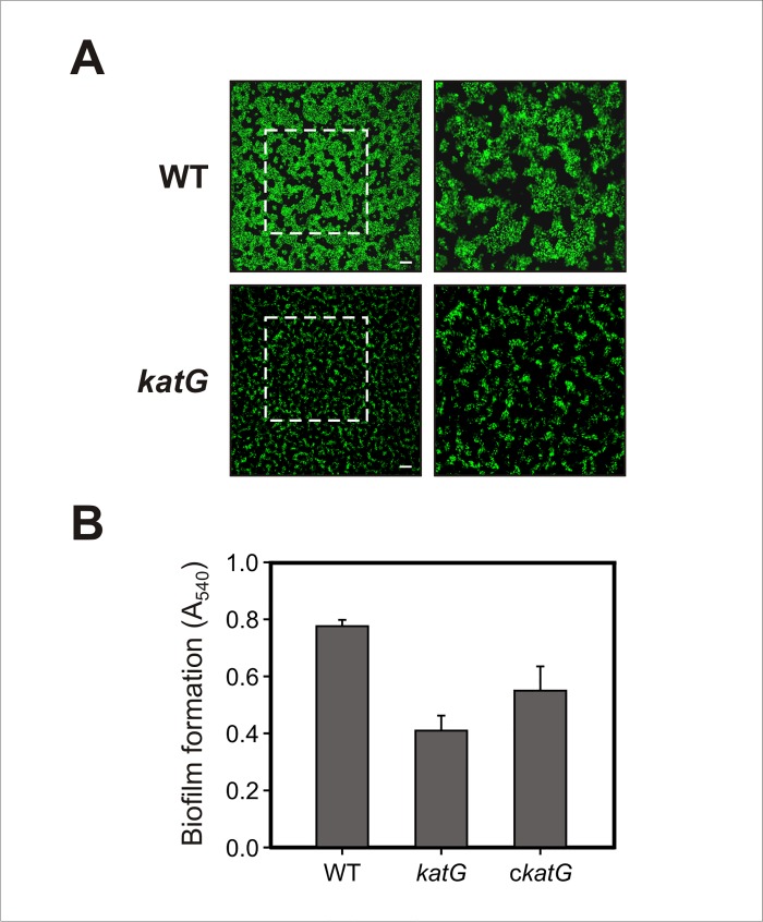
Fig 5. Effect of katG disruption on biofilm formation.
(A) GFP-labeled Xcc strains were grown on chambered cover slides and visualized under confocal laser scanning microscopy (CLSM) after 2 days of bacterial growth. Left panels show the biofilms developed at the bottom of the chambered cover slides with a magnification of 400X and right panels show a 2X zoom of the regions marked in the previous panels. Scale bars, 50 μm. (B) Xcc strains were statically grown on glass tubes for 12 days at 28°C. Biofilm formation levels on the air-liquid interface were determined by crystal violet staining. The results show the means and standard deviations of a representative experiment with triplicate samples. The experiment was repeated three times with similar results in all cases.

