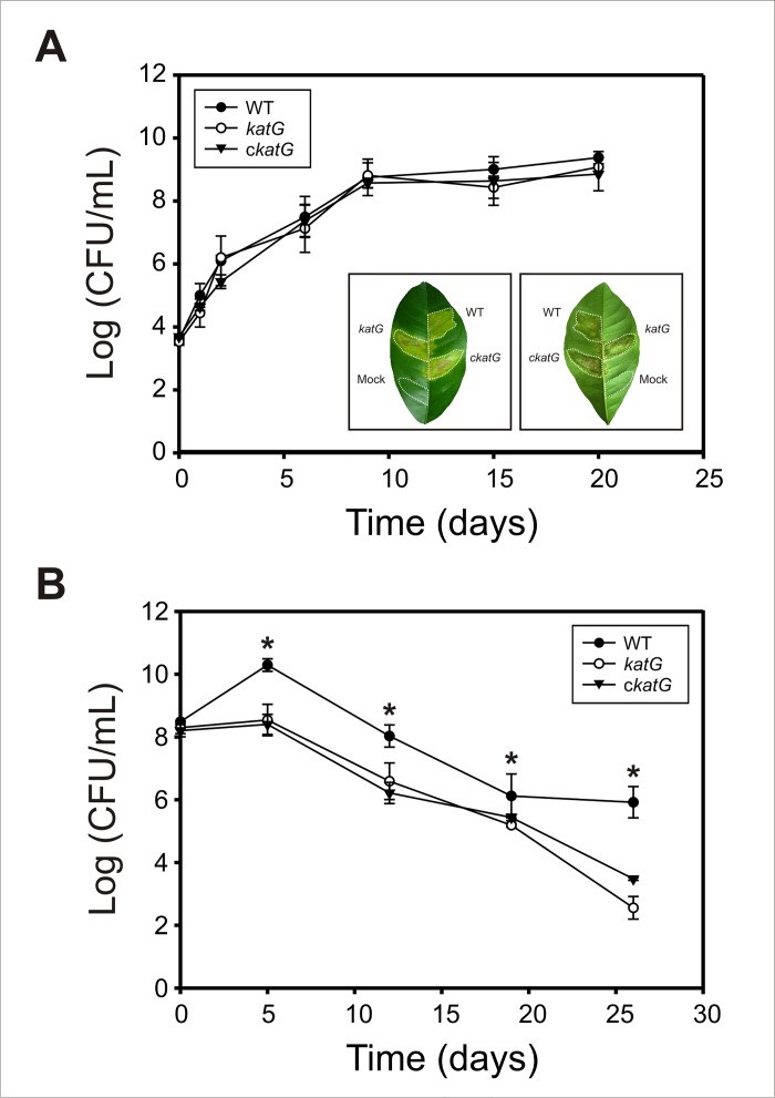
Fig 6. Pathogenicity and epiphytic fitness of XcckatG in orange plants.
(A) Growth of Xcc strains in the apoplastic space of orange leaves. Xcc WT, XcckatG and cXcckatG cells were inoculated at 105 CFU/mL in 10 mM MgCl2 into the intercellular spaces of fully expanded orange leaves. Bacterial populations in leaf tissues were determined by serial dilution and plating. A representative leaf 20 days after inoculation with the three strains is shown in the lower inset. Left panel, adaxial side; right panel, abaxial side. Dashed lines indicate the infiltrated area. (B) Epiphytic populations of Xcc strains on orange leaves. Bacterial cells were released from the leaf surface by sonication followed by dilution plating. Experiments were performed in triplicate; values are expressed as means ± standard deviations. Statistical significant differences (P < 0.05, ANOVA) between wild-type and katG strains are indicated by an asterisk.

