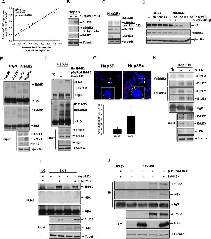
Figure 5. Overexpression of HBx enhanced the dimerization of ErbB2 and ErbB3.

A. The protein expression in human HCC cell lines was examined by Western blot and quantified by image J system. The expression level of ErbB2 and ErbB3 were normalized to its β-actin. The coefficient of determination (R2) between ErbB3 and ErbB2 expression level was analyzed by simple regression, p < 0.001. B. Hep3B cells were transfected with DsRed vector or DsRed fused ErbB3. The protein expression was determined by Western blot. C. Hep3Bx cells were infected with shRNA, the protein expression was determined by Western blot. D. Hep3Bx cells were infected with shRNA and transfected with HA-ErbB2 for 2days. The protein expression was determined by Western blot. E. Total lysate prepared from Hep3B and Hep3Bx cells were subjected to IP/Western blot analysis. F, I. and J. Hep3B (F) or HEK293T (I and J) cells were transient transfected with ErbB2, ErbB3, and vector or HBx, respectively. The total lysates were harvested for IP/Western blot with indicated antibodies. G. Hep3B and Hep3Bx were seeded for 24 hour and detected the interaction of ErbB3 and ErbB2 by using PLA kit. The red fluorescence was detected by confocal microscope. H. Hep3Bx cells were transfected with control or ErbB3 siRNA for 3 days. The total lysates were harvested for IP/Western blot with indicated antibodies.
