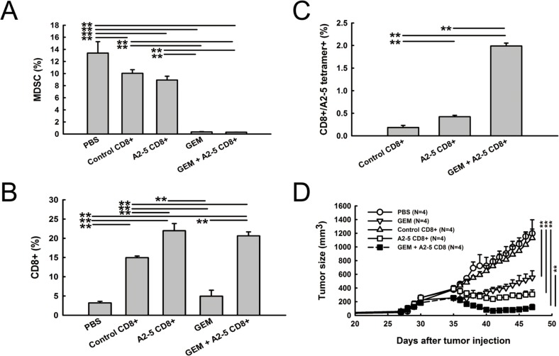
Figure 6. Reduced MDSC and increased peptide A2-5 induced CD8+ T cells were detected in xenograft tumors.
After treatment as described in Figure 5, H2981 tumors were collected on day 26 from SCID mice bearing human lung tumor xenografts. All tumors were subjected to enzymatic digestion to obtain single-cell suspensions for cell population analysis. A. The percentage of MDSC was detected with FITC-conjugated anti-GR-1 antibody and PE-conjugated anti-CD11b antibody. B. Tumor infiltrated CD8+T cells were stained with FITC-conjugated anti-CD8. C. The percentage of A2-5-HLA-A2 tetramer-binding cells was detected by flow cytometry. Error bars show mean and SD. **P < 0.01. D. H2981 cancer cells (1 × 10⁸) were subcutaneously injected in SCID mice. After 30 days, gemcitabine was i.p. injected (3mg/mouse). Isolated CD8+ T cells (1 × 10⁷) were adoptively transferred into SCID mice bearing xenograft tumors at day 35.

