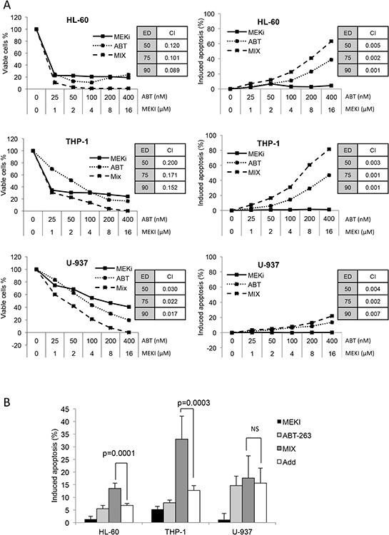
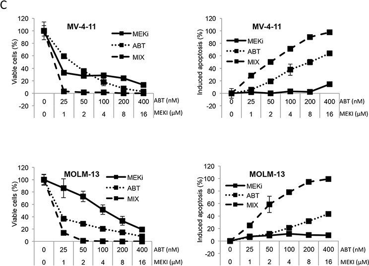
Figure 1. MEKI and ABT-263 synergize to inhibit cell proliferation and induce apoptosis in AML cell lines.


A. and C. HL-60, THP-1 or U-937 (A) and MV-4–11 or MOLM-13 (C) cells were incubated with increasing concentrations of ABT-263, MEKI or both at a constant ratio (Mix). The amount of viable cells was measured through their ATP content by chemoluminescence after a 72-h culture (left column). The percentage of apoptotic cells was determined by flow cytometry with a MMP probe after a 24-h incubation (right column). Combination indexes were calculated at ED 50, 75 and 90 using the Calcusyn software. B. HL-60, THP-1 or U-937 cells were treated for 24 h with 1 μM MEKI (black bars), 200 nM ABT-263 (light grey bars) or both (MIX, grey bars). Apoptosis was then measured by flow cytometry as described in Figure 1. Mean +/− SD of seven experiments is shown. The calculated additive effect of both drugs is also shown (white bars) and compared to the measured MIX effect using the Student paired t test. C. MV-4–11 or MOLM-13 (C) cells were incubated with increasing concentrations of ABT-263, MEKI or both at a constant ratio (Mix). The amount of viable cells was measured through their ATP content by chemoluminescence after a 72-h culture (left column). The percentage of apoptotic cells was determined by flow cytometry with a MMP probe after a 24-h incubation (right column).
