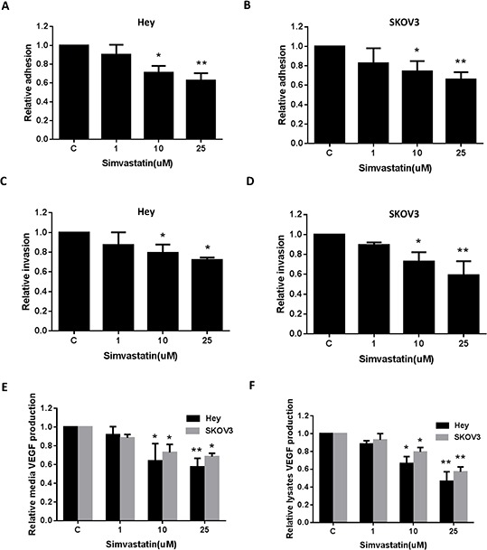
Figure 5. Simvastatin reduced on adhesion and invasion in ovarian cancer cells.

The Hey A, C. and SKOV3 B, D. cell lines were cultured for 24 h and then treated as indicated with simvastatin in a laminin coated 96 well plate for 2 h to assess adhesion or a BME coated 96 transwell plate for 24 h to assess invasion, respectively. The data represents relative inhibition in each cell line. VEGF was measured by ELISA assay in culture media E. and cell lysates F. after a 36 h exposure to simvastatin. Each experiment was performed three times. (*p < 0.05, **p < 0.01). C in graphs refers to control.
