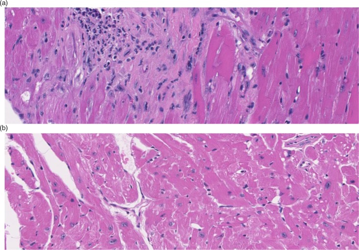
Fig. 1.
Hematoxylin- and eosin-stained heart tissue shows striking histological changes in 32-month-old CB6F1 male mice (a) compared with strain and gender-matched 8-month-old mice (b). The heart section from the old mouse shows an increased cellular infiltrate indicative of localized reactive sites, and extensive areas of fibrosis. These lesions help explain the cardiac dysfunction and increased heart weight observed in the older-aged mice.

