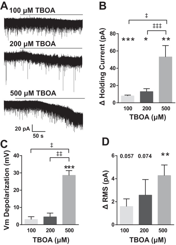
Fig. 2.

EAAT blockade depolarizes nTS neurons. A: representative traces of spontaneous activity showing the inward current produced in response to increasing concentrations of dl-threo-β-benzyloxyaspartic acid (TBOA). Cells were voltage clamped at −60 mV. B: group data showing the total change in holding current. All 3 concentrations were significantly different from aCSF baseline. Among concentrations, responses to 500 μM TBOA were significantly greater than both 100 and 200 μM. C: membrane potential (Vm) depolarized in response to TBOA although only 500 μM was significantly different from baseline. The amount of depolarization was significantly greater for 500 μM compared with either 100 or 200 μM. D: all 3 concentrations of TBOA increased root mean square (RMS) noise but only 500 μM differed significantly from baseline and 100 and 200 μM, n = 6; 500 μM, n = 7. *vs. individual baseline; ‡vs. TBOA concentration as indicated. *‡P < 0.05; **‡‡P < 0.01; ***‡‡‡P < 0.001.
