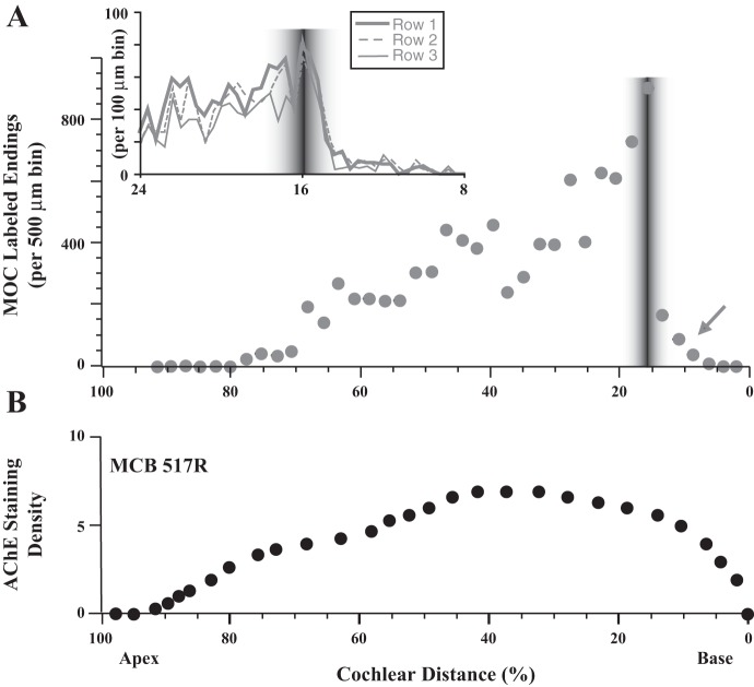
Fig. 8.
Comparison of MOC labeled endings (A) and staining for acetylcholinesterase (AChE; B). Labeling in A is the same as cases shown in Fig. 7, aligned on the basis of the middle of the ANF labeling. Inset shows an expanded view of endings tabulated separately for the OHC rows. AChE staining used a qualitative scale ranging from 0 (no staining) to 5 (darkest staining) because staining was too dense to resolve individual endings. Results for 1 cochlea are shown in B; the other 3 AChE-stained cochleas had similar patterns.

