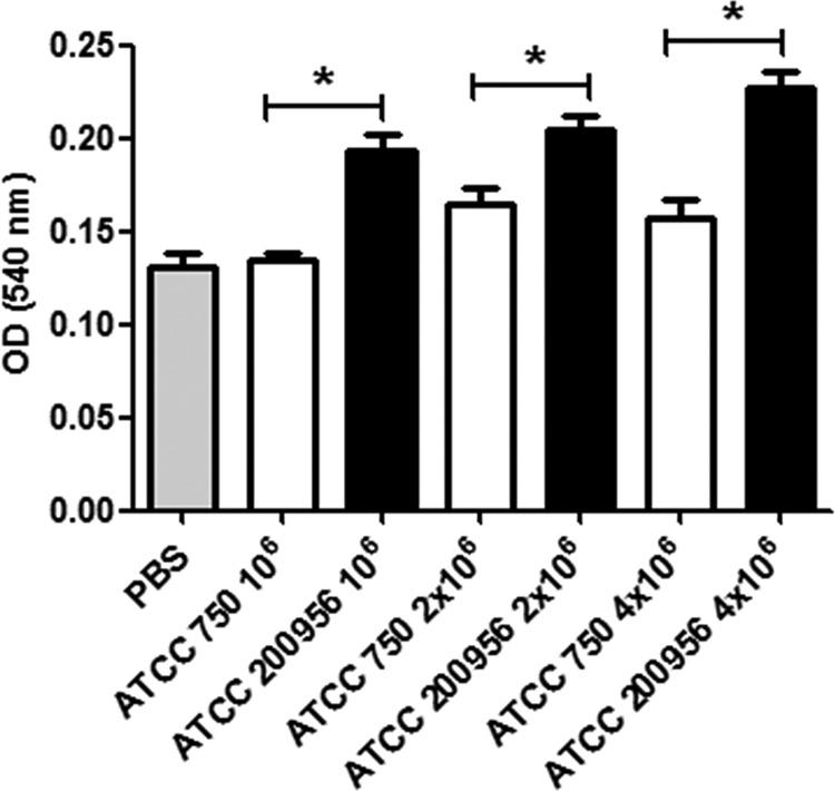
FIG 6.

Quantification of melanin in the hemolymph of G. mellonella larvae infected with C. tropicalis. Galleria mellonella larvae were infected with 106, 2 × 10⁶, or 4 × 10⁶ C. tropicalis cells from strain ATCC 750 or ATCC 200956. The larvae were incubated at 37°C, and after 1 h, the hemolymph was isolated and melanin was estimated as described in Materials and Methods. The results correspond to the average ± standard deviation of the OD values obtained from each larva in each group. The asterisks indicate significant differences (P < 0.05).
