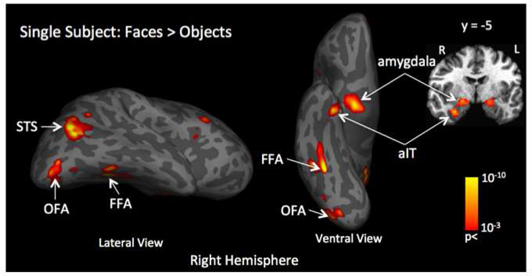
Figure 2.
Example of localization of face-selective regions by contrasting the fMRI response evoked by face stimuli compared to the response evoked by object stimuli (face > object) for a single subject. This contrast identified five face-selective regions in the right hemisphere: the dorsal-lateral part of the amygdala; anterior inferior temporal cortex - aIT; fusiform face area -FFA; posterior superior temporal sulcus - posterior STS and occipital face area – OFA. Primary visual cortex - V1 was selected as a control region of interest.

