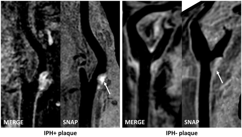
Figure 1. Plaques with and without IPH shown on isotropic three-dimensional MRI.
Left panel: Curved planar reformation (CPR) images show the left carotid artery of a 66-year old male (systolic blood pressure: 140 mmHg, diastolic blood pressure: 60 mmHg, antiplatelet use: dual therapy). Hyperintense signals on SNAP indicates IPH. Right panel: CPR images show right carotid artery of a 69-year old female (systolic blood pressure: 140 mmHg, diastolic blood pressure: 92.5 mmHg, antiplatelet use: aspirin alone).

