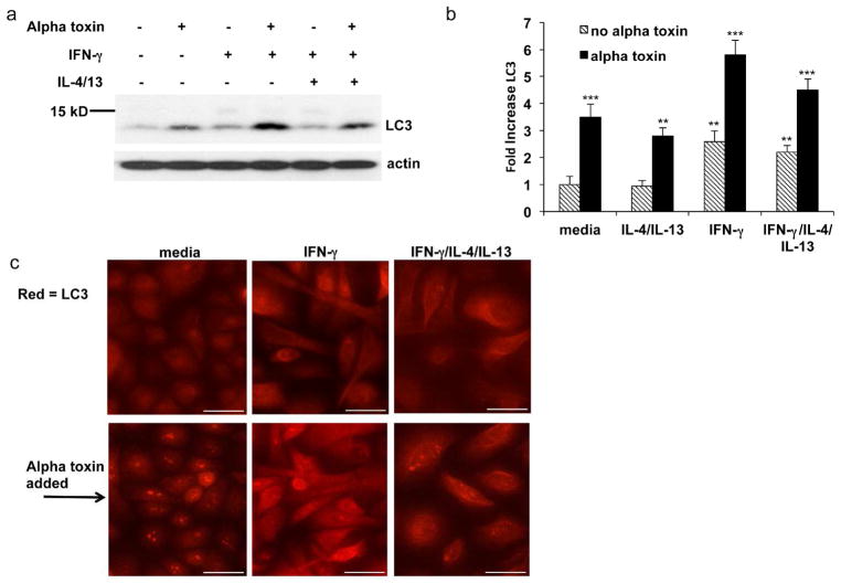
Figure 5. IFN-γ augments alpha toxin induced autophagy by increasing LC3 levels and modulating LC3 localization.
Primary keratinocytes were treated with media or the indicated cytokines for 24 hrs. The indicated cells were then treated with 100 ng/ml alpha toxin for an additional 2 hours. (a) Cells were harvested and levels of the autophagy marker, LC3, were determined by Western blot. (b) Quantitation of LC3 protein levels after pre-treatment with the indicated cytokines followed by further treatment with media or alpha toxin. (c) Immuno-fluorescence microscopy of LC3 protein. Scale bar = 50 μm. Increases in LC3 (b) are mean ± SEM, n = 3. **P<0.01; ***P<0.001 (as compared to the cells grown in medium alone).

