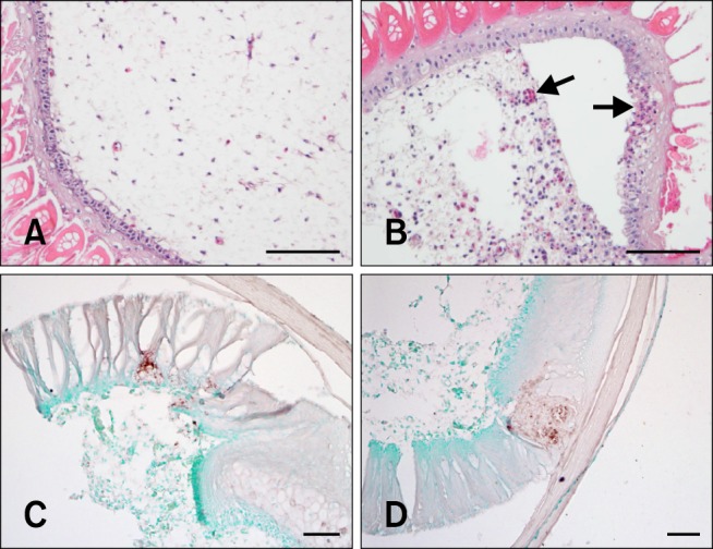
Fig. 2. Pathologic changes and immunohistochemistry for viral nucleoprotein in the feathers from vvNDV infected chickens. (A) No histological lesions in the feathers of non-infected chickens. (B) Focal necrosis of feather epidermal cells with moderate heterophilic and lymphocytic infiltration and acute heterophilic perivascular cuffing around small capillaries at the epidermal junction in the inner feather pulp from chickens 3 days post infection. (C and D) Immunohistochemistry for viral nucleoprotein of NDV. H&E stain (A and B), Immunohistochemistry stain (C and D). Scale bars = 50 µm.

