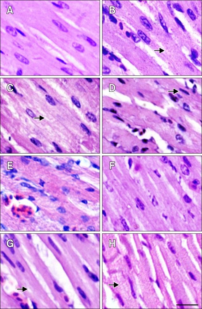
Fig. 2. Pathological changes in the myocardial fibers of tested chickens. (A–D) HS group. (E–H) ASA+HS group. (A) Heart tissue at 0 h of HS. (B) After 2 h of HS, the myocardial fibres showed the decreasing in size and granular degeneration characterised by numerous tiny granules in the cytoplasm (arrow). (C) After 5 h, vacuolar degeneration characterized by tiny water droplets in the cytoplasm, and partly dissolved muscle fibrils (arrow) was observed occasionally. (D) After 10 h, necrosis characterized by pyknosis (arrow) was evident. (E) At 0 h of HS, hemangiectasis and hyperemia were observed. (F) After 2 h, myocardial fibers sizes enlarged. (G) After 5 h, granular degeneration and vacuolar degeneration were observed occasionally (arrow). (H) After 10 h, swollen myocardial cells characterized by numerous cytoplasmic particles (arrow) were observed. H&E stain. Scale bar = 10 µm.

